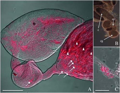
Figure 3.—
Fluorescent in situ hybridizations of W. pipientis in reproductive tissues of N. giraulti. W. pipientis (red) is shown in (A) the testes of an infected IntG12.1 male and (C) the spermatheca of an uninfected female mated to a IntG12.1 male. The ovaries of an uninfected female are also shown in B. e, mature egg at the base of a ovariole; cg, colleterial glands; sp, spermatheca. Arrows indicate W. pipientis aggregation within the testes (A) and may indicate the cellular unit transmitted to the spermatheca of uninfected females (C). Bars, 50 μm.

