
Figure 3. Down-regulation of Foxp3 expression on Tregs through OX40 costimulation and their suppressive function.
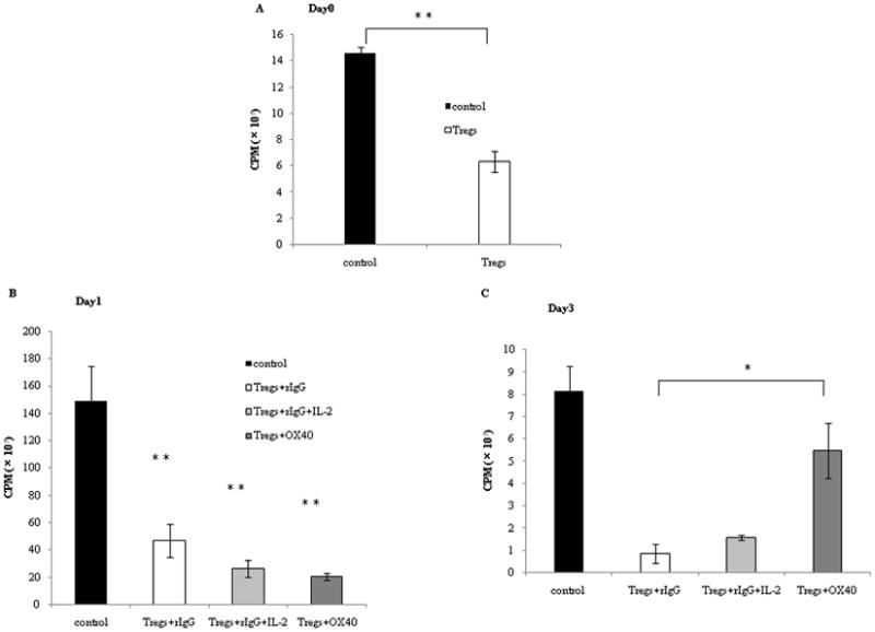
Tregs were stimulated with plate-bound anti-CD3 (5 μg/ml) in the presence of anti-OX40 mAb (5 μg/ml) or rat IgG (5 μg/ml) for 3 days.
A, B, C: the rat IgG group was divided into two groups, with or without IL-2 (10 ng/ml). Naive CD4+ T cells (2.5 ×10⁴ cells/well) were mixed with or without those Tregs which had been stimulated with rIgG or anti-OX40 mAb by the same method (2.5 × 10⁴ cells/well) and plated into 96-well tissue culture plates. The cells were incubated with irradiated APCs (2.5 ×10⁵ cells/well) and anti-CD3 (0.25μg/ml) at 37°C for 60 hours. For the last 10 hours of culture, 1 μCi [methyl-3H]Thymidine/well was added, and incorporation of thymidine was determined.
We examined the change of the suppressive function at 3 different time points: (A) before anti-CD3 stimulation, (B) day 1 and (C) day 3. “Control” indicates naïve CD4+ T cells proliferation without Tregs. In these figures, “OX40” indicates the agonistic anti-OX40 mAb. Results are from 1 representative out of 2 independent experiments. The significance of the data was evaluated with the Student’s t -test (*, p<0.05; **, p<0.01).
D: Tregs were stimulated with plate-bound anti-CD3 (5 μg/ml) in the presence of anti-OX40 mAb (5 μg/ml) or rat IgG (5 μg/ml) for 3 days. CD4+ T cells were isolated from spleens of FVB/N mice and labeled by 5μM of CFSE. CFSE-labeled CD4+ T cells (2.5 ×10⁴ cells/well) were incubated with irradiated APCs (2.5 ×10⁵ cells/well), with anti-CD3 (0.1μg/ml), and with or without non-irradiated Tregs (2.5 ×10⁴ cells/well), at 37°C for 4 days. Thereafter, dilution of CFSE gated by CD4+ T cells was visualized by flow cytometry.