
Figure 5.
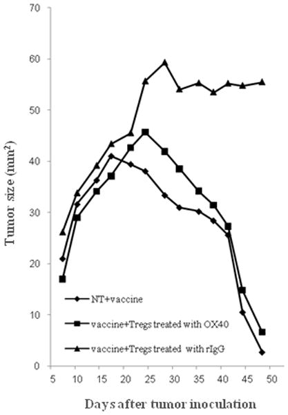
Decrease of the suppressive function of Tregs on antitumor effect through OX40 costimulation in vivo. NT2 tumor cells (5×10⁶ cells per animal) were inoculated on day −3 to FVB mice (n=20). Anti-OX40 mAb or rat IgG (300μg per animal injected i.p.) was injected on day −2 to neu-N mice (n=6 per group). On day 0, Tregs were isolated from the spleens of the injected neu-N mice using a MACS CD4+CD25+ regulatory T cell Isolation Kit. The Tregs (5×10⁵ cells/body) were adoptively transferred to the NT tumor inoculated FVB mice (n=6 per group) via their tail veins. As a control saline was injected into tail veins of FVB mice instead of adoptive transfer (n=8). 3T3 neu/GM vaccine cells (3×10⁶ total cells) were also given on day 0 to all the tumor inoculated FVB mice. Tumor size (mm2) was determined by measuring the tumor diameter along orthogonal axes. The mean tumor size is reported. “vaccine alone” indicates the tumor inoculated and vaccinated mice with saline injection. “vaccine + Tregs treated with rIgG” indicates the tumor inoculated and vaccinated mice which underwent adoptive transfer of Tregs with pretreatment of rIgG, and “vaccine + Tregs treated with OX40” indicates the tumor inoculated and vaccinated mice that underwent adoptive transfer of Tregs with pretreatment of OX40 costimulation. Statistical analysis was performed with the Students t -test.