
Figure 6. NSPCs support brain revascularization following ischemia.
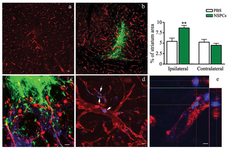
a and b) Coronal sections through the lesioned striatum of EGFP-NSPC recipients (b) or recipients of control PBS injections (a) immunostained for the endothelial cell marker, Glut-1 (red). Transplanted EGFP-NSPCs are visible as green cells. c,d,e) Double labeling of endothelial cells (Glut-1+, red) and proliferating cells (Ki67+, blue). EGFP-NSPC grafts are shown on green channel. The presence of Ki67+ nuclei within Glut-1+ blood vessels was assessed using confocal microscopy viewed in 3-dimensional (d) and orthogonal (e) image projections. The images were acquired using Zeiss LSM510- META confocal imaging system. Bars=20 μm (a-c), 10μm (d) and 2μm (e). Graph) Percentage of striatal area covered by Glut-1 immunoreactive vessels in PBS control mice (open bars) vs. EGFP-NSPC recipients (filled bars). Image analysis was performed over entire striatal area within three histological sections containing grafted cells or PBS injection sites on both lesioned (ipsilateral) and non-lesioned (contralateral) sides.