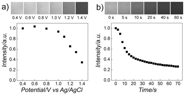
Figure 2.
Electrochemically controlled release of QDs from gold surfaces in Dulbecco’s modified Eagle’s medium (DMEM). a) Fluorescence intensity versus applied potential on a gold surface functionalized with QDs via biotin-terminated thiols (biotin-terminated tri(ethylene glycol)hexadecanethiol) (top). The fluorescence of the gold surface was measured after applying a potential for 30 s sequentially. Applying a potential larger than the critical potential (~1.0 V) induced the desorption of the SAM and thus the attached QDs and their diffusion into the bulk solution, decreasing the fluorescence signal. Corresponding fluorescence images at the labeled electrical potentials of the gold surface in gray scale are shown at the top of the panel, showing gradual loss of the fluorescence. b) Fluorescence intensity versus time curve of a gold surface functionalized with QDs at a constant applied potential of 1.4V versus Ag/AgCl wire in DMEM. The fluorescence intensity decreased as the SAM and the attached QDs were desorbed and diffused into the bulk solution. Corresponding fluorescence images at the indicated time are shown at the top of the panel.

