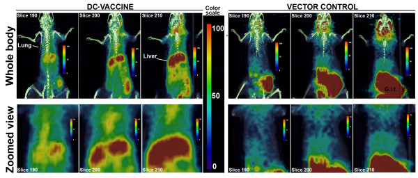
Figure 10.
Fused positron emission tomography (PET) and computed tomography (CT) images of BALB/c mice intranasally instilled with homologous primary DCs expressing HSV1-TK. The primary DCs were either co-transfected with pVR1012-Ag2/PRA-cDNA and pVR1012-TK (labeled as DC-vaccine), or with vector plasmid DNA (labeled as Vector Control). The PET-CT imaging was performed after 7 days of cells administration. About 2 h prior to imaging, 18F-FIAU (a substrate for HSV1-TK) was intravenously injected. Images are from one representative mouse from each group. Six mice were included per group in three different experiments. In the second panels of each group, the whole body images were zoomed to get closer view of 18F-FIAU accumulation (DC-migration) in thoracic area. A significant accumulation of 18F-FIAU was observed in lung and liver of mouse injected with DCs expressing TK. Overall, the mice receiving DC-vaccine retained more 18F-FIAU radioactivity than the Control mice. G.i.t (gastrointestinal tract).

