Figure 5.
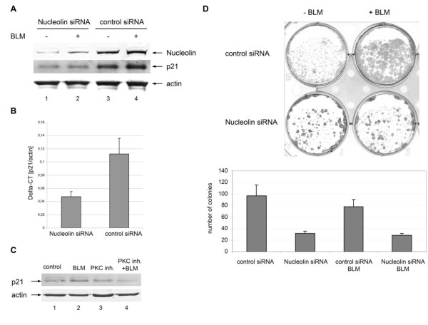
Depletion of nucleolin induced downregulation of p21 and impeded cell proliferation. (A) Protein extract of human HaCaT cells transfected with specific nucleolin siRNA (lanes 1-2) or unspecific control siRNA (lanes 3-4) which were treated with BLM for 30 min (lanes 2 and 4) or untreated cells (lanes 1 and 3) were subjected to immunoblotting against nucleolin and p21 using specific antibodies. As a control for equal protein loading corresponding actin levels are shown by immunoblot. (B) HaCaT keratinocytes transfected with specific nucleolin siRNA or unspecific control siRNA were cultured for 72 h and treated for 1hour with BLM (12.5 μg/ml). Then total RNA was extracted from the cells and p21 gene expression analysis was performed. The CT values of p21 were normalized to actin. Two independent experiments were performed and data are displayed as mean values (±SD). (C) HaCaT cells treated with BLM, the myristoylated PKC inhibitor, or both BLM and PKC inhibitor, and, as a control, untreated cells were lysed and protein extracts were subjected to immunoblotting using a specific antibody against p21. As a control for equal protein loading corresponding actin levels are shown by immunoblot. (D) Decreased proliferation capacity of nucleolin-depleted HaCaT cells. For assessment of the proliferation capacity of HaCaT keratinocytes, colony formation assays were carried out. HaCaT cells were transfected with a specific nucleolin siRNA or, as a control, with unspecific control siRNA, treated with BLM 72 h after transfection and grown for further 7 d (+BLM). As a control, transfected cells grown without BLM treatment (-BLM). For quantification, the colonies in 3 representative areas (1.0 cm diameter) of 6-well dishes with transfected HaCaT cells with (+BLM) or without (-BLM) BLM treatment were counted. Data are displayed as mean values (±SD).

