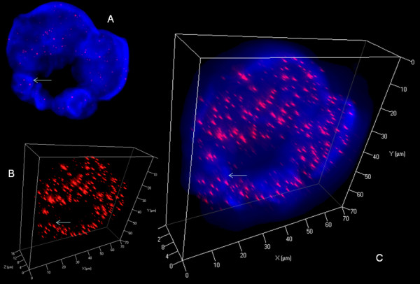
Figure 6.

3D nuclear staining of telomeres and DNA in U-HO1 RS-cells. A. Representative 2D Z-stack image no. 36 out of 80 shows ring-like multinuclear (blue) RS-cell composed of at least four nuclei of variable size. Arrow identifies a telomere group also shown in B and C. B. 3D reconstruction in surface mode reveals abundant short and very short telomeres (red). C. Combined 3D nuclear staining confirms ring-like nuclear configuration.
