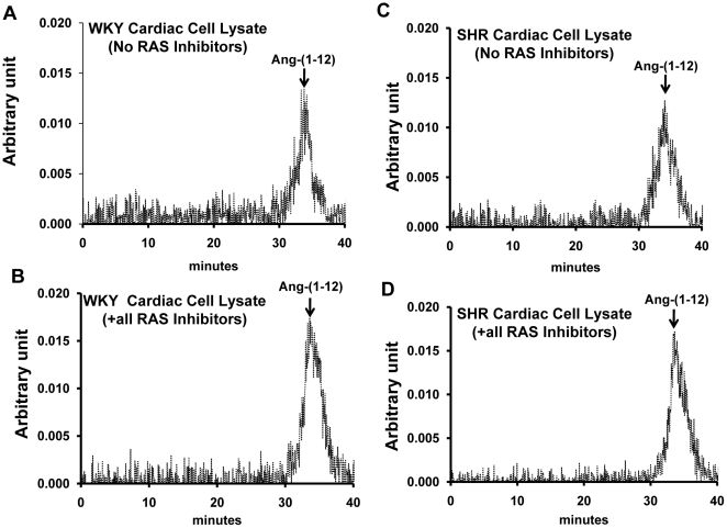
Figure 2. Effect of inhibitors on Ang-(1–12) metabolism in cardiac myocytes.
Characterization by high pressure liquid chromatography of cellular 125I-Ang-(1–12) products internalized by 24-h serum deprived cultured neonatal WKY (A and B) and SHR (C and D) cardiac myocytes in the presence of all inhibitors cocktail (containing lisinopril, SCH39370, MLN-4760, chymostatin, amastatin, bestatin, benzyl succinate and PCMB) and in the absence of RAS inhibitors (containing only amastatin, bestatin, benzyl succinate & PCMB) each added at a dose of 10 µM. Before adding the 125I-Ang-(1–12), the cultured cardiac myocytes were pre-incubated with inhibitors for 15 min at 37°C. The arrow indicates the peak area of 125I-Ang-(1–12).

