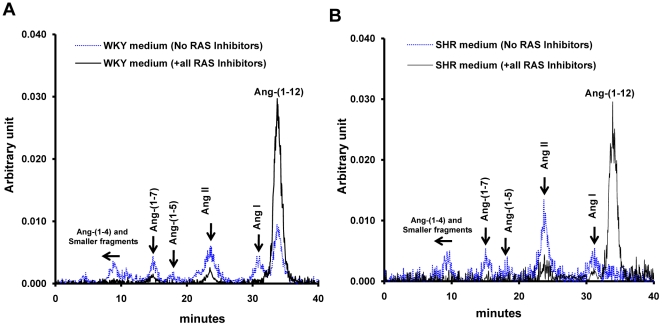
Figure 3. Patterns of Ang-(1–12) metabolism in the medium collected from cardiac myocytes.
HPLC characterization of 125I-Ang products generated in the medium of cultured neonatal WKY (A) and SHR (B) cardiac myocytes exposed to 125I-Ang-(1–12) at 37°C for 60 min in the presence (solid line) and absence (dotted line) of the RAS inhibitors cocktail. Other conditions as described in Figure 2.

