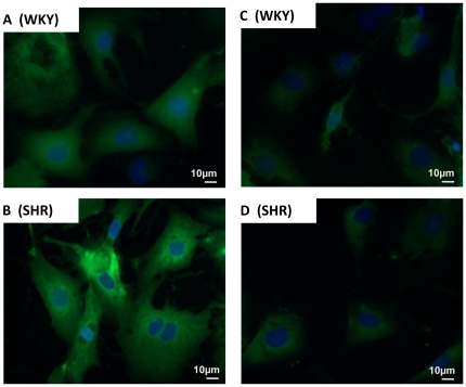
Figure 5. Characteristics of Ang-(1–12) expression in cultured myocytes.
Representative examples of fluorescent photomicrographs of Ang-(1–12) immunoreactivity of the cultured myocytes (maintained for 48 hours in serum-deprived medium) from WKY (A and C; top panel) and SHR (B and D; bottom panel) with protein A purified Ang-(1–12) polyclonal antibody (1∶100 dilution). (A and B) represents the fluorescent staining of endogenous Ang-(1–12) with antibody while (C and D) illustrates the blocking of fluorescent staining of cardiac myocytes after preadsorption of the antibody with 10 µM of synthetic rat Ang-(1–12) peptide. Magnification, X400.

