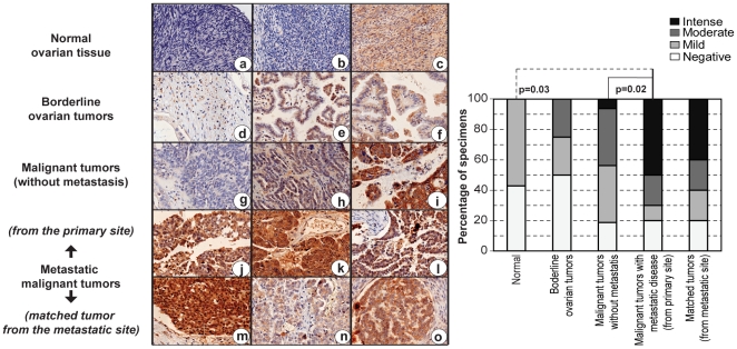
Figure 7. 14-3-3 σ expression and ovarian tumor metastasis.
Immunohistochemical analysis of 14-3-3 σ in ovarian tissues show negative (hematoxylin stained blue nucleus) to diffuse staining pattern for 14-3-3 σ (brown) in normal ovarian tissues (a–c), and a moderate increase in the staining intensity localized to the cytoplasm is observed in the borderline ovarian tumors (d–f). In the malignant tumors without any metastatic disease at diagnosis, 14-3-3 σ expression was either absent (g), or stained at moderate to intense levels (h–i), with occasional nuclear staining (i). Intense nuclear and cytoplasmic staining for 14-3-3 σ was observed in ovarian tumors with metastatic disease, obtained from the primary site of the disease, and a moderate to intense staining for 14-3-3 σ in the cytoplasm or both nucleus and cytoplasm of the corresponding tumors obtained from the metastatic site was observed [site of metastasis - (m) appendix, (n) lymph node and (o) omentum]. The quantitative relationship between 14-3-3 σ expression and various stages of ovarian cancer progression is represented in the bar graph, and the statistical analysis for correlation of expression with pathological grades were determined by a Fisher’s exact test.

