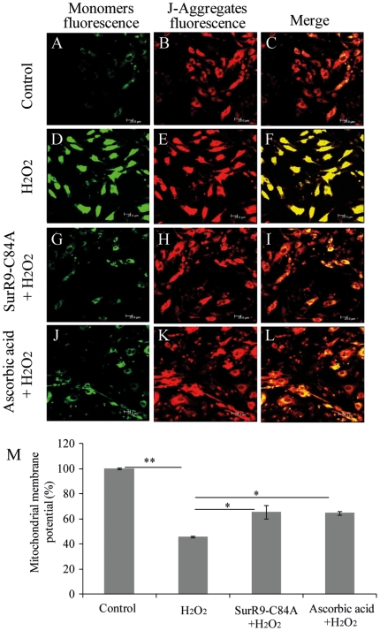
Figure 3. SurR9-C84A prevents mitochondrial depolarization.
SK-N-SH cells were differentiated with 20 µM retinoic acid for 10 days. Differentiated media were replaced with growth media and cells were pre-treated with 75 µg/ml of SurR9-C84A or ascorbic acid for 24 hr followed by treatment with 300 µM of H2O2 for 24 hr. At the end of incubation mitochondrial membrane depolarization was qualified and quantified with MitoLight Mitochondrial kit using both techniques of (A) confocal microscopy and (B) spectrofluorometery (see material and method). Green fluorescence (detection of monomers) indicates the presence of depolarized mitochondria (apoptotic cells). Red fluorescence (J-aggregates) indicates the functional and polarized mitochondria. Values are presented as a percentage of increase in mitochondrial depolarization. Data are representative of at least three independent experiments and expressed as mean±SEM; *P<0.05, **P<0.01.

