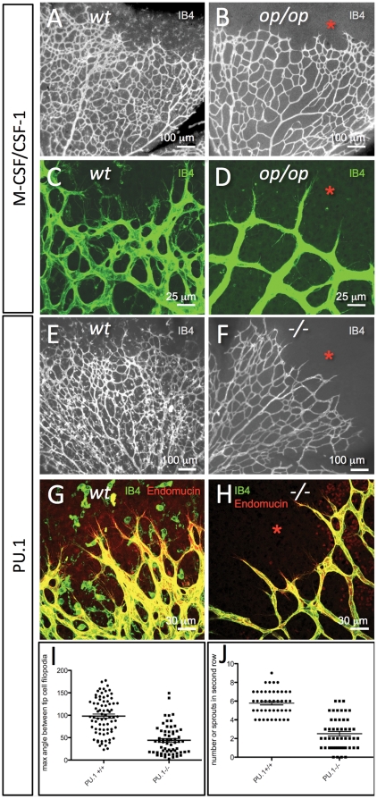
Figure 2. Absence of microglial cells correlates with morphological changes in tip-cells and in the developing retinal vascular plexus.
(A–D) Retinal vascular plexus morphology as depicted by IB4-staining in P6 mice of wildtype (A,C) or csf-1op/op genotype (B,D). Note the complete absence of IB4-stained microglial cells in the retina ahead of the vascular plexus (asterisks), and the difference in vascular density. Note also that the tip-cells and their filopodia in the csf-1op/op retinas (D) are more uniformly radial in their orientation than the corresponding tip-cells of the control (C). (E–H) Retinal vascular plexus morphology as depicted by IB4/Endomucin staining in P6 mice of wildtype (E,G) or Pu.1-/- genotype (F,H). Note the absence of microglial cells (asterisks), the sparser developing vascular plexus and a preferential radial orientation of tip-cell filopodia in the mutants (F,H). (I, J) The maximum angle between the filopodia protruding from a single tip cell is reduced in Pu.1-/- mice (I, n = 76, p<0.0001), and less tip-cells and filopodia were observed in the second row of branches, i.e. at a more central location in the retina of Pu.1-/- mice (J, n = 50, p<0.0001), suggesting that microglia promotes further angiogenic sprouting at this location.

