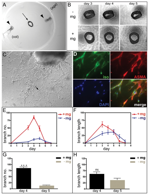
Figure 3. Microglia stimulate angiogenesis.
(A) An aortic ring explant (arrow) embedded between two disks of collagen type Ι (col) and surrounded by a cylinder of agarose type VII (aga). Microglia cells in suspension were injected between the discs at two locations (indicated by arrowheads). (B) Phase-contrast images of aortic rings explants from one mouse cultured in the absence (-mg) or in the presence (+mg) of microglia. The images show the same aortic ring explants after three, four and five days in culture. (C) An inverted microscope image showing an aortic ring culture with neovessels at day 4 (arrow points to a vessel). (D) Whole mount immuno-staining of an aortic ring co-cultured with microglia. The aortic ring explant was stained with IB4 (green) for endothelial cells, mouse monoclonal anti-α-smooth muscle actin antibody (red) for pericytes, and DAPI for nucleic acids (blue), and their merged image is shown. (E,F) Numbers and lengths of the vascular branches in 24 aortic ring explants (from four mice) cultured with or without microglia were determined each day for 7 days. The diagrams indicate the mean relative values (the ratio between the particular value and the highest value in the specific series) for number (E) and length (F) of the vascular branches of four independent series of experiments. (G and H) Mean relative values obtained on the day of maximum angiogenic response (day four for cultures in the presence of microglia and at day five for cultures in the absence of microglia) in terms of number (G) and length (H) of the vessels in the four independent experiments above. Bars indicate standard errors of the means. The angiogenic response of aortic rings in terms of branch number was significantly increased in the presence of microglia (***p<0.001), whereas the difference in response in terms of branch length was not statistically significant (ns).

