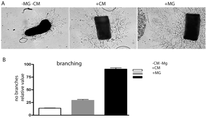
Figure 7. The angiogenic response of aortic rings in culture is significantly higher in the presence of activated microglial cells compared with in conditioned medium only.
(A) Phase-contrast images of cultured mouse aortic ring explants with standard culture medium (left), microglia conditioned medium (middle) and with microglia (right). The images show aortic rings at day five of culturing. (B) Diagram showing the mean number of vascular branches in 12 aortic rings for each experimental condition. Conditioned medium was obtained from the same number of microglia as was applied in the co-culture experiment. Bars indicate standard errors of the means. The angiogenic response of aortic rings cultured in the presence of microglia cells was significantly higher than that of aorta rings grown in conditioned medium (p<0.001).

