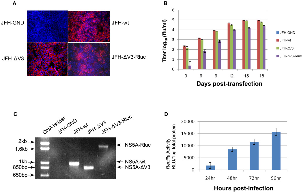
Figure 3. Infectivity assay of virus particles produced following RNA transfection of cells.
Panel A, detection of HCV replication by NS5A immunofluorescence assays (IFA) following infection of naïve cells (see Materials and Methods). Huh 7.5 cells were infected with the supernatant collected at 15 days after transfection with JFH-GND, JFH-wt, JFH-ΔV3, and JFH-ΔV3-Rluc RNA. Panel B, Production of infectious HCV particles in cell culture supernatants following transfection with viral RNA (see Materials and Methods). The viral titer is expressed as focus-forming units per ml of supernatant (ffu/ml) as determined by the average number of NS5A-positive foci detected by immunofluorescence for NS5A. Assays were done in triplicate in 96-well plates and performed two times. The data are presented as mean ± standard deviation (n=6). Panel C, Detection of HCV virion RNA in the culture medium by RT-PCR spanning the NS5A V3 region (see Materials and Methods). The DNA products were analyzed by 1% agarose gel electrophoresis. Experiments were performed three times and a representative experiment is shown. Panel D, Renilla luciferase activity was measured in the Huh 7.5 cells following infection with the JFH-ΔV3-Rluc virus at a multiplicity of infection (moi) of 0.1 (see Materials and Methods). Assays were done in triplicate three times and the data are shown as mean ± standard deviation (n=9).

