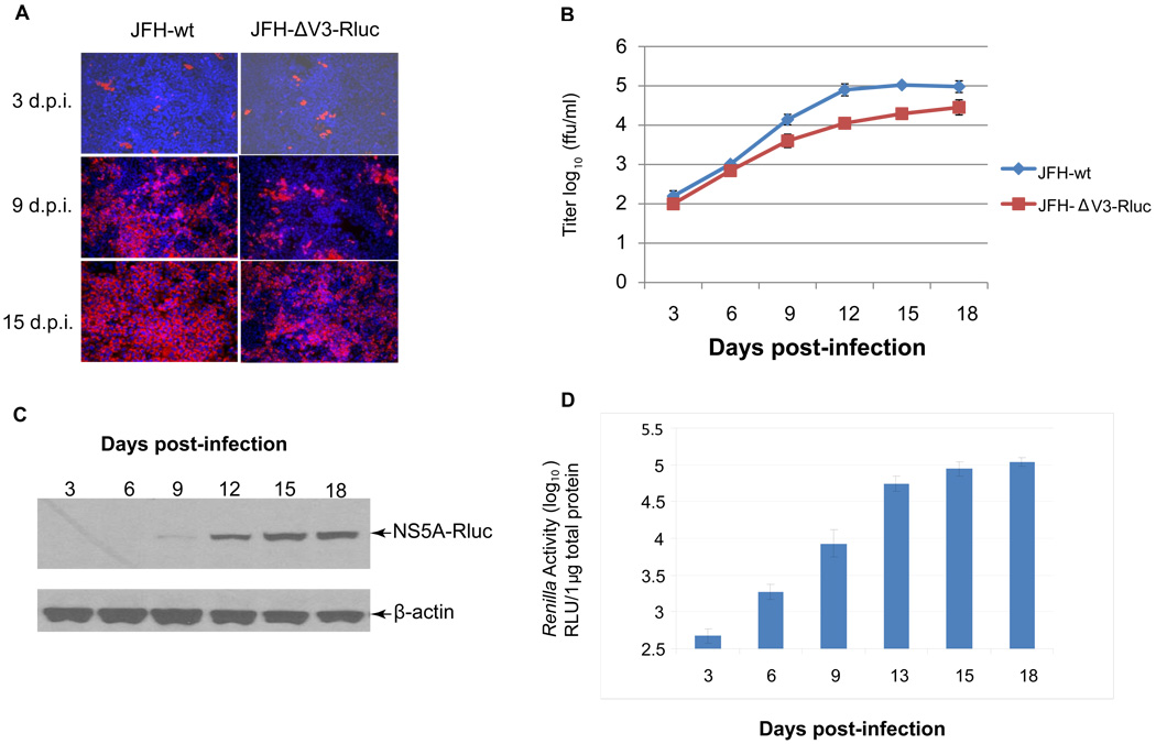
Figure 4. Kinetics of virus production following infection of naïve Huh 7.5 cells with JFH-ΔV3-Rluc or JFH-wt virus.
Panel A, naïve Huh 7.5 cells were infected with JFH-wt and JFH-ΔV3-Rluc supernatants at an moi of 0.01. Cells were passaged every three days and analyzed at the indicated times for NS5A expression by immunofluorescence (red). Nuclei were counterstained using DAPI (blue). Panel B, virus titers in cell culture supernatants collected at the indicated times post-infection were measured by determining focus-forming units with NS5A immunofluorescence assays. Assays were done in triplicate, performed twice and data presented as mean ± standard deviation (n=6). Panel C, Detection of the NS5A-Rluc fusion protein in serially passaged cells by Western blotting with anti-Rluc. Experiments were performed twice and a representative result is shown (see Materials and Methods). Panel D, Renilla luciferase activity was measured at the indicated times post-inoculation with the JFH-ΔV3-Rluc virus. Assays were done in triplicate, performed twice and data shown as mean ± standard deviation (n=6).

