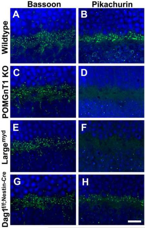
Figure 3.
Diminished pikachurin expression in the outer plexiform layer of the mutant mouse retinas.
Retinal sections were immunofluorescence stained with antibodies against Bassoon (A, C, E, and G) and pikachurin (B, D, F, and H). (A and B) Wildtype retina. Bassoon and pikachurin immunofluorescence were observed at the ribbon synapses in the outer plexiform layer. (C and D) POMGnT1 knockout retina. While ribbon synapses were immunoreactive to anti-Bassoon, immunoreactivity to anti-pikachurin was lost. (E and F) Largemyd retina. Pikachurin immunoreactivity at the ribbon synapse was lost. (G and H) Dag1f/f;Nestin-Cre(+) retina. While no noticeable changes were observed for the density of Bassoon immunoreactive ribbon synapses, the density of pikachurin-positive puncta was reduced. Each pikachurin-positive puncta showed similar fluorescence intensity to the wildtype. Scale bar in H: 10 μm.

