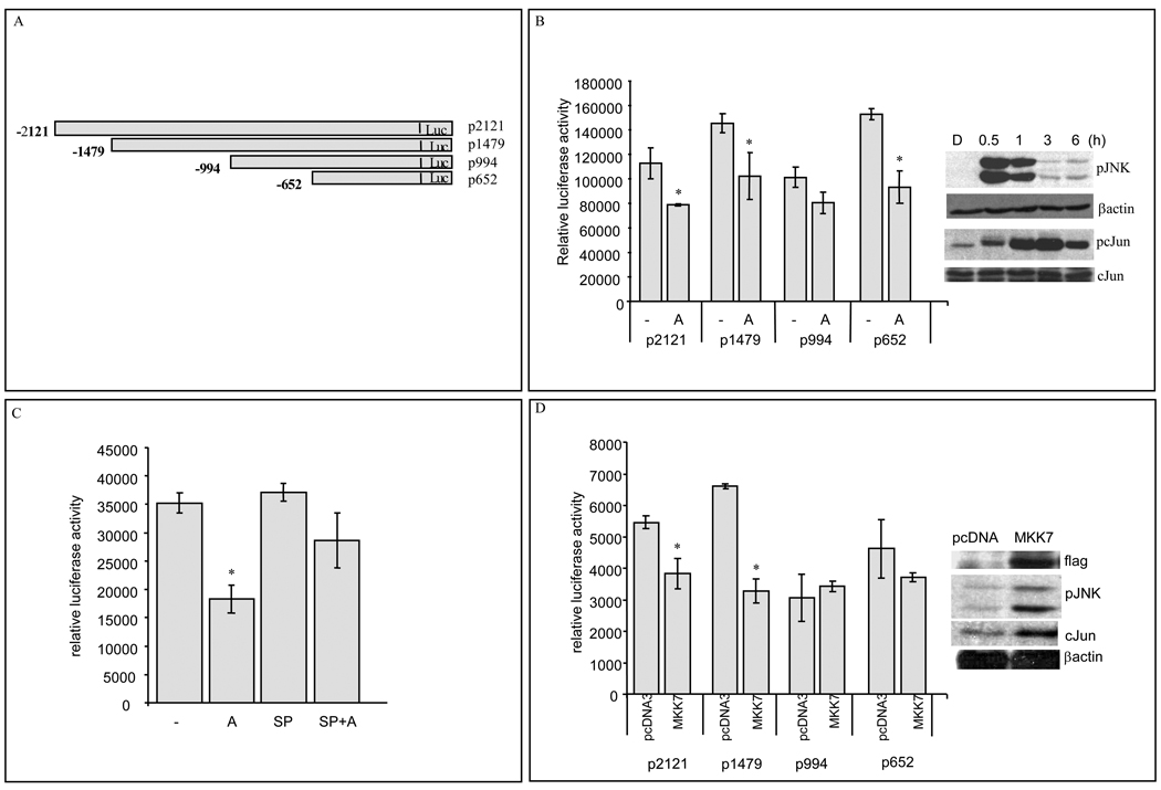
Figure 3. JNK activation decreases mENT1 promoter activity.
Data points represent sample means ± S.D. of data (A) mENT1 promoter picture (B) Relative promoter activity in anisomycin treated cells. HEK293 cells transfected with different mENT1 promoter fragments were treated with anisomycin (A) 50ng/ml for 6h. (Representative experiment, n=6, *p<0.05).). Right panel: Immunoblot analysis for pJNK, pc-Jun (Ser73), total c-Jun and β-actin from HEK293 cells treated with anisomycin 50ng/ml for the times indicated. (C) mENT1 pLuc-2121 promoter fragment activity in HEK293 cells treated with anisomycin (A) 50ng/ml, 10µM SP600125 (SP) or combination of SP and anisomycin (SP+A) for 6h. (Representative experiment, n=6, *p<0.05). (D) Promoter activity in flag-MKK7 expressing HEK293 cells cotransfected with different mENT1 luciferase-coupled promoter fragments. (Representative experiment, n=3, *p<0.05). Right panel: Immunoblot analysis for flag-MKK7, pJNK, total c-Jun and β-actin from HEK293 cells transfected with pcDNA3 or MKK7 expression vector.

