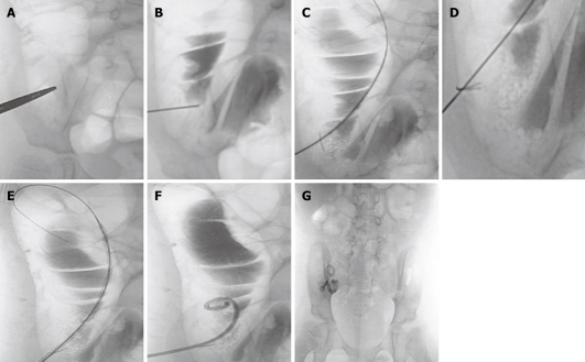
Figure 1.
Procedure of percutaneous cecostomy. A: Skin entrance site is localized over the air-filled distended cecum; B: After needle insertion into the cecum, contrast is injected to confirm its location within the cecal lumen; C: Guide wire insertion into the distended right side of the colon; D: T-fastener deployed to fix the anterior wall of the cecum to the abdominal wall; E: The insertion tract is dilated before placement of the cecostomy tube; F: Locking pigtail catheter is inserted into the cecum; G; After 6 wk, the Dawson Mueller catheter is exchanged for the permanent Chait Trapdoor cecostomy catheter.

