
Figure 1. Nucleotide and predicted amino acid sequence of the lamprey neurofilament protein L-NFL.
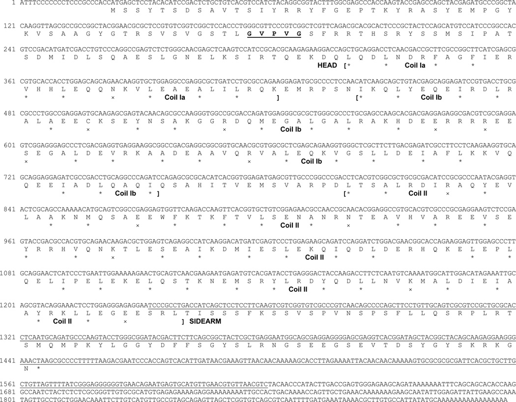
Translation was begun at the first in-frame methionine of the longest open reading frame and terminated by the stop codon “TAA”. Nucleotide numbers are indicated at the beginning of each DNA sequence line. The protein sequence before the first bracket is the head region and that after the last bracket is the sidearm (tail) region. Protein sequence between the first and the last bracket is rod region, which consists of 3 coils (coil Ia, Ib, and II). In each coil, stars (*) mark hydrophobic residues at the first and fourth amino acid of putative heptad repeats, while exes (x) mark charged residues. The underlined region is the nucleotide sequence used for Northern blotting or in situ hybridization. The amino acids “GVPVG” in bold and underlined in the head region represents a sequence unique to L-NFL as compared with other NF-Ls listed in Table 1A.