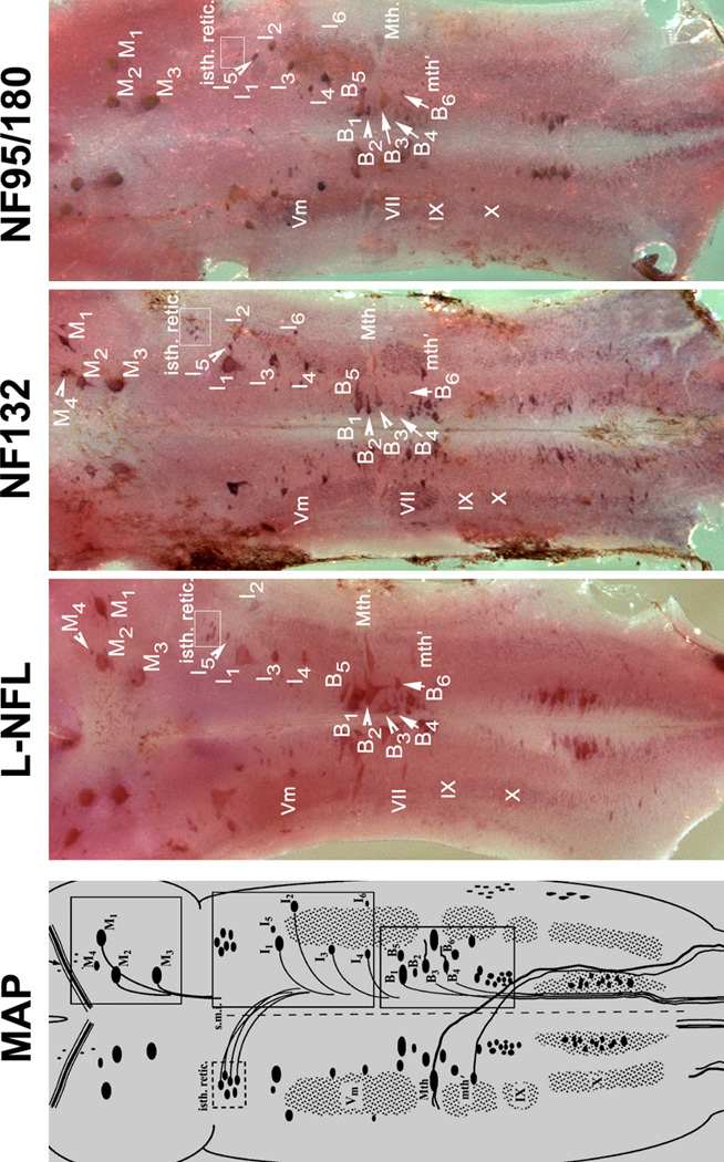
Figure 10. Expression patterns for lamprey NF mRNAs in the lamprey brain.

MAP: Schematic drawing of mature (5-year-old) larval sea lamprey brain stem, showing major anatomical features and the locations of identified neurons and the neuron groups. The view is from the dorsal surface after removal of the mesencephalic and rhombencephalic choroid plexus, transection of the cerebrotectal commissure and obex, and lateral reflection of the alar plate. The identified spinal-projecting neurons include M1-4, I1-6, B1-6, the Mauthner (Mth) and auxiliary Mauthner (Mth’) neurons, the isthmic reticulospinal (isth.retic.), and medial inferior reticulospinal (m.i.r.) cell groups, some cranial nerve nuclei [Vm (trigeminal motor nucleus), VII, IX and X] (Reprinted with permission from Jacobs et al., 1997, J Neurosci 17:5206–5220, ©1997 by the Society for Neuroscience). Three boxes on the right are the locations of 3 identified neuron groups (M-, I-, and B-groups), Mauthner (Mth) and auxiliary Mauthner (mth’) neurons. L-NFL, NF132, and NF95/180 are wholemount in situ hybridizations of lamprey brain stem with corresponding probes. Animals used were large lavae, approximately 4 years old with body lengths 130-, 135-, and 135-mm, respectively. Comparison of regions within boxes are given in Figure 11.
