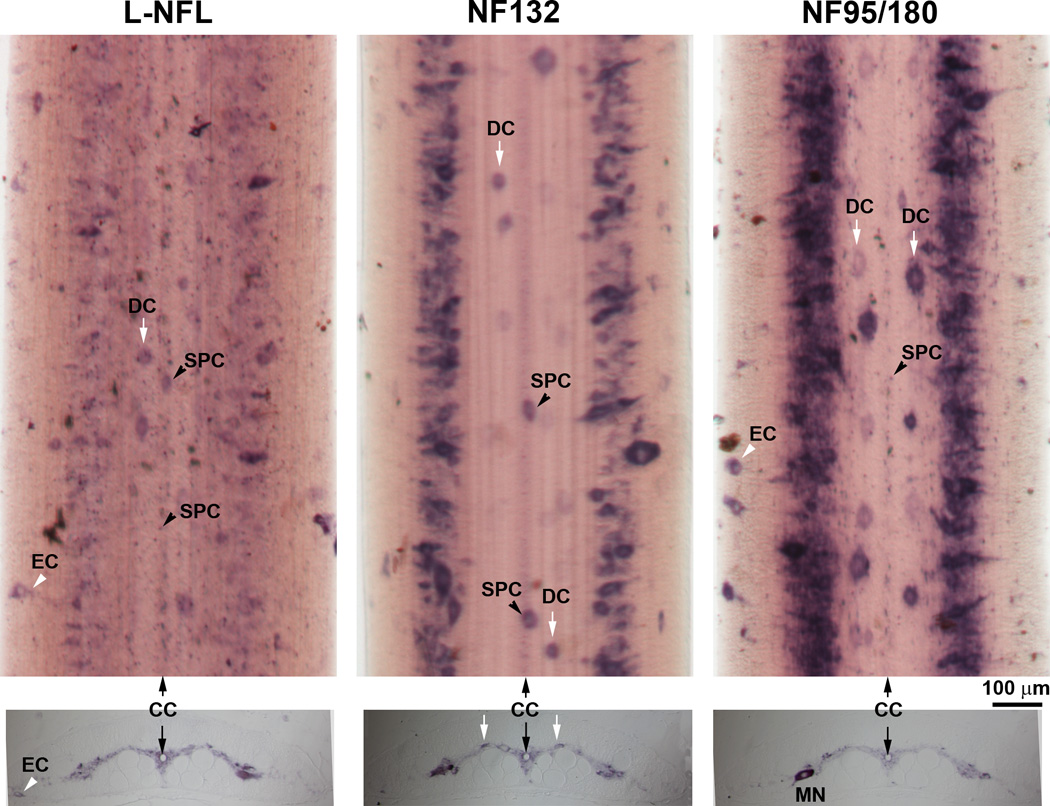
Figure 12. Cellular localization of NFs in the spinal cord.

Upper panel: In situ hybridization in spinal cord wholemounts showed a high level of NF95/180 mRNA expression along the lateral gray matter. L-NFL was expressed relatively evenly and weakly along the entire spinal cord. Several neuron types can be identified by their morphology and location, including: dorsal cells (DC); subependymal cells (SPC) surrounding the central canal (CC), and edge cells (EC). Note: NF132 labeled only large and medium sized cells. Lower panel: In situ hybridization in transverse sections from a 12 cm long larva (approximately 3–4 yrs old). All neurons are localized in the gray matter except for ECs, which sit along the edge of the spinal cord. MN: possible motor neuron, based on size, location and the ventrally projecting axon hillock, which is faintly labeled.
