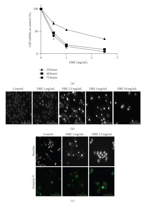
Figure 1.
Determining the sensitivity of A375 human melanoma cells to DRE (a) Effect of DRE on cell viability. A375 human melanoma cells were seeded on 96-well plates (~1000 cells/well) and treated at the indicated concentrations for 24, 48, and 72 hours. The WST-1 dye was added to each well after every treatment period and incubated, as described in Section 2. Absorbances were read at 450 nm. (b) Induction of apoptosis by DRE. Typical apoptotic morphology was observed in A375 cells treated with DRE (0–10 mg/mL concentrations) for 48 hours. Cells were stained with Hoechst 33342 dye, before images were taken on a fluorescence microscope. Brightly stained, condensed bodies indicate apoptotic nuclei. (c) Confirmation of apoptosis by Annexin-V binding assay. Cells treated at the effective and subeffective doses of 2.5 and 1 mg/mL, respectively, for 48 hours were stained with Annexin-V Alexa Fluor (green) following which cells were imaged on a fluorescence microscope.

