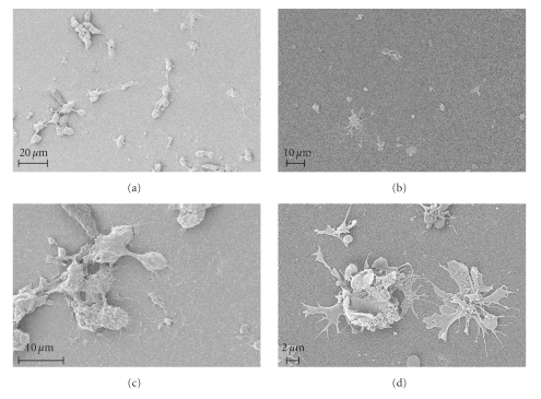
Figure 4.
SEM micrographs of monocytes/macrophages after 24 h of incubation on nanoporous alumina. MM on the 200 nm-pore membranes ((b) and (d)) show clear signs of activation (rough plasmalemma and extended filipodia), while cells on the 20 nm-pore membranes ((a) and (c)) appear more round shaped with little membrane ruffling and no established filipodia extensions. A higher tendency toward cell clustering and fusion was also observed on the 20 nm-pore alumina (c).

