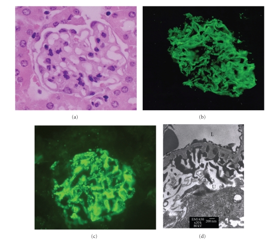
Figure 2.
Renal histopathology in mice with MN. Histopathology revealed findings characteristic of diffuse basement membrane thickening, as observed in the (a) hematoxylin and eosin staining, (b) positive granular immunofluorescent staining for IgG, (c) positive granular immunofluorescent staining for C3, and (d) subepithelial deposition (asterisk). NC: normal control; MN: membranous nephropathy; G: glomerular basement membrane; L: lumen of capillary; U: urinary space.

