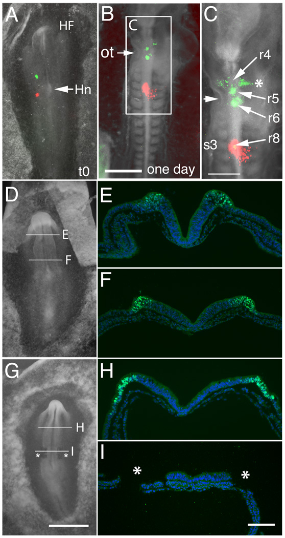
Figure 2.
Fate map analysis of normal and ablated embryos. (A–C) Dye-labeling of control embryos reveals that presumptive cardiac neural crest lies in the neural folds at the rostral margin of forming somite 1 (DiO, green) and caudal margin of forming somite 3 (DiI, red) in a representative embryo at stage 7 (A, t=0min). (B) Twenty-four hours after labeling. (C) Enlargement of “B”. By stage 12/13, the green neural fold injection originally placed at somite 1 level now labels the neural tube/neural crest at the mid-otic level (ot, arrow; rhombomeres 5/6 area); the red neural fold injection spot originally placed at somite 3 level remains at the same somite 3 level (rhombomere 8). The neural fold cells at the level of somite 1 at stage 7 translocate rostrally and the neural fold undergoes extension, resulting in a forward shift of the neural folds of ~100 mm. The DiO-positive cells in the r4 stream (asterisk) come from r5. The two midline DiO spots are at the level of r5 and r6. (D–I) Expression of Pax7, a marker of the forming dorsal neural folds, at stage 7 in control and ablated embryos. (D) Whole mount of a stage 7 control embryo. (E) Representative section showing Pax7 expression (green) in the neural folds at the level of the midbrain. DAPI (blue) stains cell nuclei. (F) Pax7 expression in the forming neural folds at the level of the caudal hindbrain (which contains the presumptive cardiac neural crest). (G) Whole mount of a stage 7 embryo after bilateral ablation of the cardiac neural folds from level of somite 1–3 (asterisks). (H) Pax7 expression in a similar embryo, as in (G), in the neural folds at the level of the intact midbrain. (I) Absence of Pax7 expression confirms the removal of the cardiac neural folds at the level of ablation (asterisks; region of ablated folds).
Abbreviations: HF- head fold; Hn-Hensen’s node; ot-otic cup; r4, r5 r6 and r8-Rhombomeres 4, 5, 6 and 8.
Scale bars. G: 1mm, in panels A, D, G. B: 500µm. C: 300µm. I: 100µm, in panels E, F, H, I.

