Figure 4.
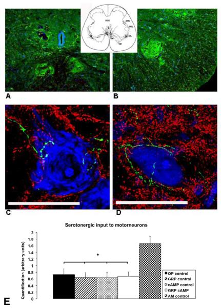
Location of dorsomedial (A) and dorsolateral nuclei (B) in the lumbosacral spinal cord, examples of motor neurons located in the dorsomedial or dorsolateral nuclei (C and D), and quantification of serotonin in close proximity to these motor neurons (E). Sections in A and B are stained for serotonin (green) and cell nuclei (blue; TOTO-3 cyanine dimer nucleic acid stain, Invitrogen Corporation, Carlsbad, CA, USA; 2mM) by immunohistochemistry. In A the 2 dorsomedial nuclei are shown left and right from midline just ventral to the central canal. B shows the right dorsolateral nucleus in the ventral horn of the lumbosacral spinal cord. Sections in C and D are 20 μm thick and stained for serotonin (green), synaptophysin (red), and DNA/RNA (TOTO-3) by immunohistochemistry. The extensive dendritic field with synapses in these nuclei and the serotonergic input (green) to the motor neurons is shown here. Scale bar (C and D) = 50 μm. In E the quantification of serotonin in close proximity to motor neurons is shown in arbitrary units for all groups of rats. ✦: significantly different from AM control.

