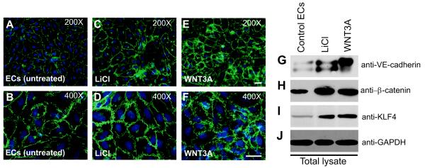
Figure 1. WNT3A induces the expression of VE-cadherin.
A-F, Untreated or ECs treated with LiCl (20 ng/mL) or with recombinant WNT3A (50 ng/mL) for 3 d and stained for VE-cadherin. Representative images of control and treated ECs at 200× (A, C, E) and 400× (B, D, F) magnification. For additional images, see Figure S2. G-J) Cell extracts were analyzed by Western blot (WB) for VE-cadherin, β-catenin, KLF4, and GAPDH. Results are representative of at least three separate experiments.

