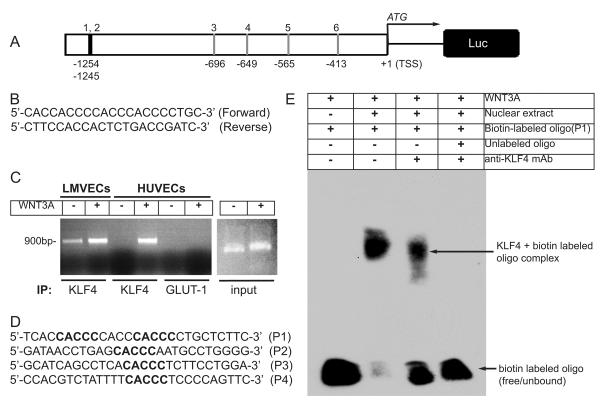
Figure 3. KLF4 binds to VE-cadherin promoter.
A, Schematic of human VE-cadherin promoter −1.3 kb upstream of transcription start site (TSS). Potential KLF4 binding (CACCC) sites are indicated. B, Sequence of human VE-cadherin primer pair used for ChIP experiments. C, HLMVECs and HUVECs were grown in complete media, and left untreated (-) or treated for 3 days with WNT3A. ChIP assay was performed with indicated antibodies. PCR product of VE-cadherin promoter using input chromatin. D, Biotin-labeled oligonucleotide probes (P1-P4) containing CACCC sites used for EMSA. E, Representative image of EMSA blot. Probe-1 (P1) of KLF4 site from VE-cadherin-promoter was incubated with nuclear extracts prepared from ECs treated with WNT3A or by pre-incubation of nuclear extract with anti-KLF4 antibody in the presence or absence of cold-unlabeled oligonucleotide. Results are representative of at least three separate experiments.

