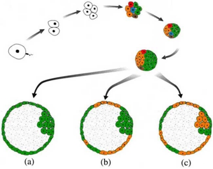
Fig. (1).
The development of a human embryo to the blastocyst stage. The different colors at the eight cell stage represent mosaicism of normal blastomeres (green) and blastomeres carrying mitotically derived aneuploidies and mitotic structural aberrations (orange, red, blue and brown). When the embryo reaches blastocyst stage, the aberrant cells can be lost due to negative selection (a); they can segregate to the trophectoderm only, leading to confined placental mosaicism (b); or they can be found in both the inner cell mass and the trophectoderm resulting in an embryo that is affected in certain tissues (c).

