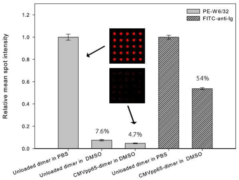
Figure 2.
Effect of dimethyl sulfoxide (DMSO) on the binding of anti-HLA (clone W6/32) and anti-Ig antibodies binding to microarray spots printed with pHLA A2-Ig dimers. From left to right: PE-labeled W6/32 (20 μg/ml) contacted with spots printed with 0.1mg/ml unloaded pHLA A2-Ig dimer in PBS solution, in 0.33% (v/v) DMSO/PBS solution, and with 66.7μg/ml CMVpp65-loaded pHLA A2-Ig dimer in 0.33% (v/v) DMSO/PBS solution, and FITC-labeled anti-Igλ (20 μg/ml) contacted with spots printed with 0.43mg/ml unloaded pHLA A2-Ig dimer in PBS solution and with 276 μg/ml CMVpp65-loaded pHLA A2-Ig dimer in 1.4% (v/v) DMSO/PBS solution. Mean spot fluorescence intensities are normalized to the mean spot fluorescence intensities for the unloaded dimer in PBS solution for each antibody staining, with the percentages shown above the bars corresponding to these relative mean spots intensities. Insets: scanned fluorescence images of subarrays for the two printing solutions designated by the arrows.

