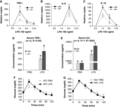
Figure 3.
Effect of FoxO1 on Tlr4 signalling in vivo. (A–C) BMDMs derived from FoxO1+/− or WT mice were overnight starved in 0.1% BSA-containing medium, and treated with 100 ng/ml LPS. Cells were subsequently harvested at the indicated time points. mRNA expression of TNFα (A), IL-6 (B) and IL-1β (C) were quantitated. Asterisks indicate statistical significant difference at each corresponding time point. (D, E) Circulating plasma levels of TNFα (D) and IL-6 (E) 3 h post-LPS injection were measured. Data are presented as the average±s.d. Letters above the bars show statistical groups (ANOVA, P<0.05). (F, G) Following 6 h fast, WT (F) and FoxO1+/− (G) mice were injected with LPS (1 mg/kg) or vehicle 1 h before i.p. injection of glucose (1 mg/kg). Blood glucose levels were measured during a 2-h GTT. Asterisk indicates a significantly higher peak glucose level at 15 min in WT mice (n=4, P=0.039).

