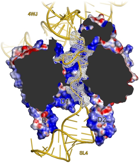
Figure 4.
Threading of the snRNA through the Sm ring. Cut-away view into the Sm ring showing the displacement of the single-stranded RNA region towards the D1-D2 sector. Sm proteins are shown in surface representation with the electrostatic surface potential in the inner pore of the Sm ring. Blue, positive charge; red, negative charge. Selected elements are labelled and coloured as in Figure 1A. 4WJ, four-way junction. Grey mesh, Fo−Fc ‘omit' electron density (with the RNA entry, Sm site and RNA exit omitted) contoured at the 2.5 σ level. Rotated 150° about the y axis compared with Figure 1A.

