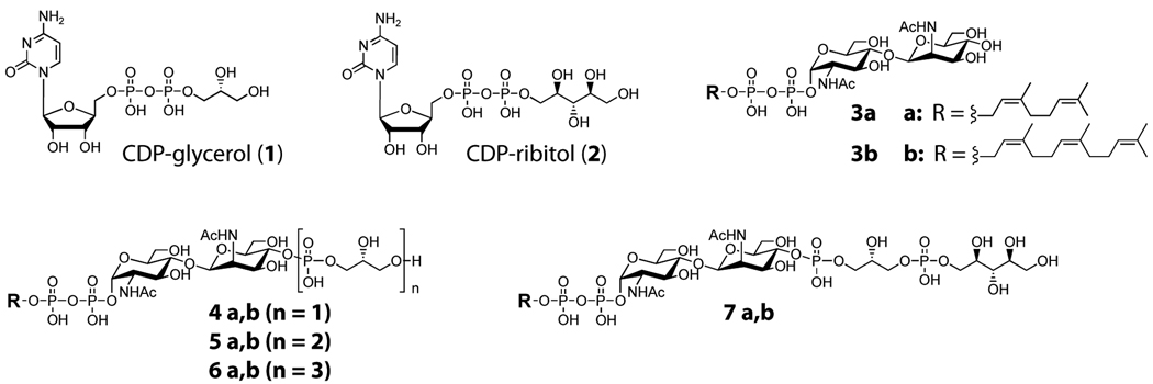
Figure 2. Structures of WTA substrates and intermediates.
The structures of the CDP-glycerol (1) and CDP-ribitol (2) donor and lipid acceptor substrates 3, 4, 5, 6, and 7. The natural acceptor substrates contain an undecaprenyl chain. Substrates having the “a” suffix contain a neryl chain and were used for LC/MS analysis; substrates having the “b” suffix contain a farnesyl chain and were used for PAGE analysis. Synthesis of substrates and incorporation of [14C] radiolabels for PAGE analysis were carried out as described previously (Brown et al., 2008).

