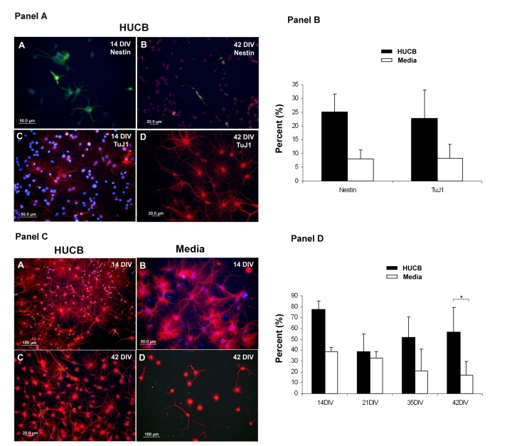
Fig. 7.
The expression of neural antigens on aging hippocampal neurons. Panel A. Expression of the stem cell (Nestin) and immature neuronal (TuJ1) antigens. (A, B) Several Nestin+ cells (green) were detected in the HUCB treated hippocampal cells at A) 14 DIV and B) 42 DIV. Similarly, a number of TuJ1+ cells (red) were observed in the HUCB treated hippocampal cultures at 14 DIV (C) and 42 DIV (D). DAPI staining was used as a counterstain to visualize nuclei (blue). Scale bar = 50 μm in (A) and (C), and 20 μm in (B) and (D). Panel B. Percentage of total cells that were Nestin and TuJ1 positive after 42 DIV. Panel C. The mature neuronal marker MAP2 is expressed on aging hippocampal neurons in culture. (A, C) show that the majority of HUCB treated hippocampal neurons expressed MAP2 (red) and had extremely rich neurite outgrowth on days 14 (A) and 42 (C). (B, D) MAP2+ hippocampal neurons (red) in the non-treated group at 14 DIV (B) and 42 DIV (D). MAP2+ cells in this group were similar to those in the treated group in the early stage of culture (14 DIV). However, the number of MAP2+ cells dramatically declined and most of them lost their processes by 42 DIV. Scale bar = 100 μm in (A) and (D), 50 μm in (B) and 20 μm in (C). Panel D. Percentage of MAP2+ hippocampal cells in culture over time. * p < 0.05.

