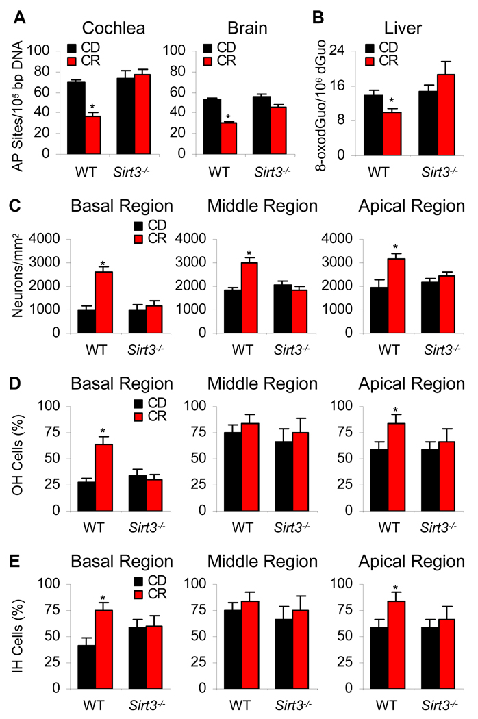
Figure 2. CR Reduces Oxidative DNA Damage and Increases Cell Survival in the Cochleae from WT Mice, But not from Sirt3−/− Mice.
(A) Oxidative damage to DNA (apurinic/apyrimidinic sites) was measured in the cochlea and neocortex from control diet and calorie restricted WT and Sirt3−/− mice at 12 months of age (n = 4–5). AP sites= apurinic/apyrimidinic sites. *Significantly different from 12-month-old WT mice (P < 0.05).
(B) Oxidative damage to DNA (8-oxodGuo) was measured in the liver from control diet and calorie restricted WT and Sirt3−/− mice at 12 months of age (n = 4–5).
(C) Neuron survival (neuron density) of basal, middle, and apical cochlear regions was measured from control diet and calorie restricted WT and Sirt3−/− mice at 12 months of age (n = 4–5).
(D) OH (outer hair) cell survival (%) of basal, middle, and apical cochlear regions was measured from control diet and calorie restricted WT and Sirt3−/− mice at 12 months of age (n = 4–5).
(E) IH (inner hair) cell survival (%) of basal, middle, and apical cochlear regions was measured from control diet and calorie restricted WT and Sirt3−/− mice at 12 months of age (n = 4–5). Data are means ± SEM. See also Figure 1B–M.

