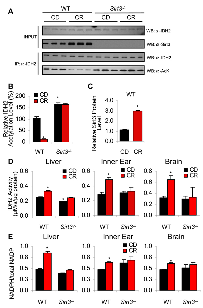
Figure 4. Sirt3 Increases Idh2 Activity and NADPH Levels in Mitochondria by Decreasing the Acetylation State of Idh2 During CR.
(A) Top panels: Western blot analysis of Sirt3 and Idh2 levels in the liver from 5-month-old WT or Sirt3−/− fed either control or calorie restricted diet. Lower panels: Endogenous acetylated Idh2 was isolated by immunoprecipitation with anti-Idh2 antibody followed by western blotting with anti-acetyl-lysine antibody (n = 3).
(B–C) Quantification of the amounts of total Idh2 acetylation (B) and Sirt3 protein (C) from (A). Western blot was normalized with Idh2 levels or Sirt3 levels quantified and analyzed by Image software (n = 3).
(D) Idh2 activities were measured in the liver, inner ear (cochlea), and brain (neocortex) from control diet and calorie restricted WT and Sirt3−/− mice at 5 months of age (n = 3–5).
(E) Ratios of NADPH:total NADP (NADP+ + NADPH) were measured in the liver, inner ear, and brain (neocortex) from control diet and caloric restricted WT and Sirt3−/− mice at 5 months of age (n = 3–5). *Significantly different from control diet fed WT mice (P < 0.05). Data are means ± SEM.

