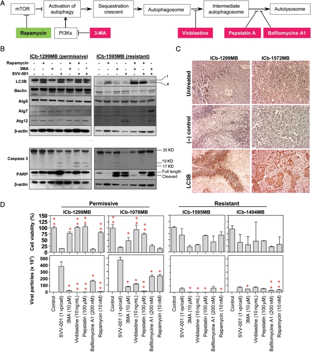
Fig. 5.
Induction of autophagy and apoptosis by SVV-001. (A) Overview of the steps activating autophagy and the specific activator. (B) Changes of autophagy (upper panel) and apoptosis (lower panel) genes induced by SVV-001 in vitro. Primary cultured cells from the permissive model ICb-1299MB and the resistant model ICb-1595MB were treated with SVV-001 (MOI 25) for 24 hours before being subjected to western hybridization. The autophagy inducer rapamycin (10 nM) and inhibitor 3MA (10 µM) were also included as controls. (C) IHC detection of LC3B expression in vivo in the 2 permissive xenograft mouse models 48 hours after SVV-001 single tail vein injection. (D) Impact of autophagy inhibitors (3MA, vinblastine, pepstatin, and bafilomycin A1) and activator (rapamycin) on the cell killing (upper panel) and extracellular viral production (lower panel). Cell viabilities in cells treated with combined drug (inhibitor or activator) and SVV-001 were normalized to those treated with drug only and presented as percentages. Treatment with autophagy inhibitors led to increased cell viability in the permissive models (upper panel) and decreased extracellular viral production in both the permissive and resistant models (*P< .05 and **P< .01 when compared with SVV-001 only) (lower panel).

