Fig. 3.
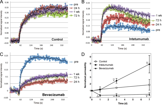
Effect of intetumumab and bevacizumab on vascular permeability. Rats with intracerebral LX-1 SCLC xenografts underwent serial DCE-MRI with GBCA. Changes in normalized signal intensity are shown in (A) control untreated rat; (B) rat treated with intetumumab 30 mg/kg i.v.; (C) rat treated with bevacizumab 45 mg/kg i.v. Scans were obtained before treatment (pre; blue) and 24 hours (red), 72 hours (green), and 1 week (purple) after treatment. (D) The time-intensity mean and the standard error are indicated for n = 4–6 rats per time point, showing increased permeability after intetumumab and decreased permeability after bevacizumab. In comparison with baseline, *P < .05 and **P < .001; in comparison with control at each time point, •P < .05 and ••P < .001.

