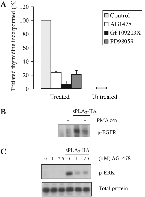
Fig. 5.
sPLA2-IIA–induced proliferation in 1321N1 cells and sPLA2-IIA signaling induced in MCF7 cells. (A) Proliferation was measured as described in the Methods section. Serum-starved 1321N1 cells were stimulated with sPLA2-IIA for 24 hours after preincubation for 30 minutes with the indicated inhibitors (1-μM AG1478, 1-μM GF109203X, or 25-μM PD98059) or vehicle. [3H]Thymidine incorporated in sPLA2-IIA–stimulated cells was taken as 100%, and the rest of the values were related to this. Bars represent the means ± S.E. of 3 independent experiments performed in triplicate. (B and C) Serum-starved MCF7 cells were stimulated with sPLA2-IIA for 5 minutes after an overnight incubation with 1-μM PMA (B) or a 30-minute incubation with AG1478 (C) at the indicated doses. Total lysates were subjected to SDS-PAGE and incubated with phospho-EGFR (B) or phospho-ERK (C).

