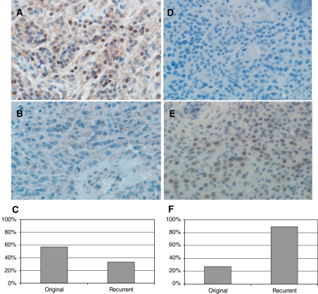
Fig. 3.
Immunohistochemistry results of 2 genes differentially expressed in original and recurrent tumors. Photomicrograph of meningioma samples with (A) positive and (B) negative expression of LMO4 (original magnification, ×400). (C) Percentage of cases with LMO4-positive expression. Photomicrograph of tumor tissue with (D) negative and (E) positive expression of HIST1H1C (original magnification, ×400). (F) Percentage of cases with HIST1H1C-positive expression.

