Fig. 6.
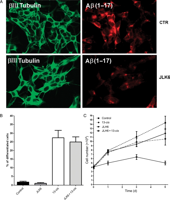
Neuroblastoma cell proliferation and differentiation analysis after JLK6 treatment. SH-SY5Y neuroblastoma cells stably transfected with APP 751 wild type (SH-SY5Y-APPwt) were treated for 5 days with 1 µM JLK6 GSI, with 1 µM 13-cis RA, and with the combination of the 2 compounds. (A) Determination of Aβ peptide levels in control and JLK6-treated cells. The βIII tubulin antibody (green) identified neuronal cells. Anti-Aβ 6E10 antibody (red) was used to detect amino acids 1–17 of Aβ peptide. (B) The Histogram represents the percentage of differentiated cells. (C) Cell growth determination by a Trypan blue exclusion test at 1, 3, and 5 days after treatment.

