Fig 5.

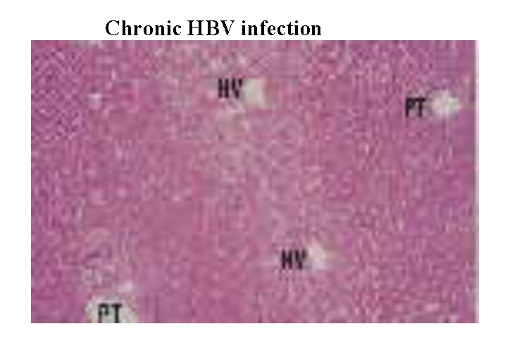
Section of liver showing normal architecture with regularly spaced portal tract (PT) and hepatic venues (HV). Haematoxylin and eosin stain; magnification approximately x35. (16)

Section of liver showing normal architecture with regularly spaced portal tract (PT) and hepatic venues (HV). Haematoxylin and eosin stain; magnification approximately x35. (16)