Fig. 10.

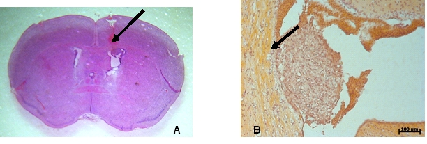
Macroscopic (10A) and microscopic (10 B–inverted image) aspect of the glioblastoma xenograft at 7 days postinoculation (arrow). Colored with hematoxilin–eosin.

Macroscopic (10A) and microscopic (10 B–inverted image) aspect of the glioblastoma xenograft at 7 days postinoculation (arrow). Colored with hematoxilin–eosin.