Fig. 11.

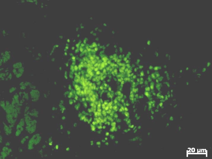
The fluorescence microscopic aspect of the GFP transfected glioblastoma cells at 3 days post–inoculation. Computerized system acquisition has been used for this image

The fluorescence microscopic aspect of the GFP transfected glioblastoma cells at 3 days post–inoculation. Computerized system acquisition has been used for this image