
Fig 9.
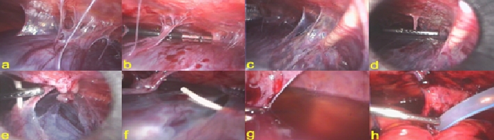
CSF pseudocyst. a. general view of the pseudocyst; b. adhesiolysis; c. distal catheter entering the pseudocyst; d. e. adhesiolysis of the distal catheter; f. distal catheter freed from the pseudocyst; g. partially evacuated pseudocyst; h. drainage of the pouch of Douglas.