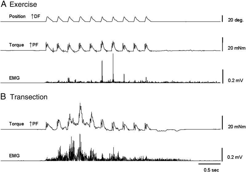
Figure 1. Stretch reflex recordings showing the SR wind-up protocol at a 0.25 sec interval.
Representative data is shown at 49 days post spinal cord transection (Tx) for a Tx only animal and an animal in the Ex group. Ankle position, ankle torque and rectified gastrocnemius EMG are plotted across time. A) After 6 weeks of daily exercise, animals demonstrated SR activity (see EMG) but responses did not wind up with repeated stretches. B) Tx animals showed a strong SR wind-up of both the EMG and torque responses to repeated stretches with a peak during the 4th repetition. DF – dorsiflexion, PF – plantar flexion.

