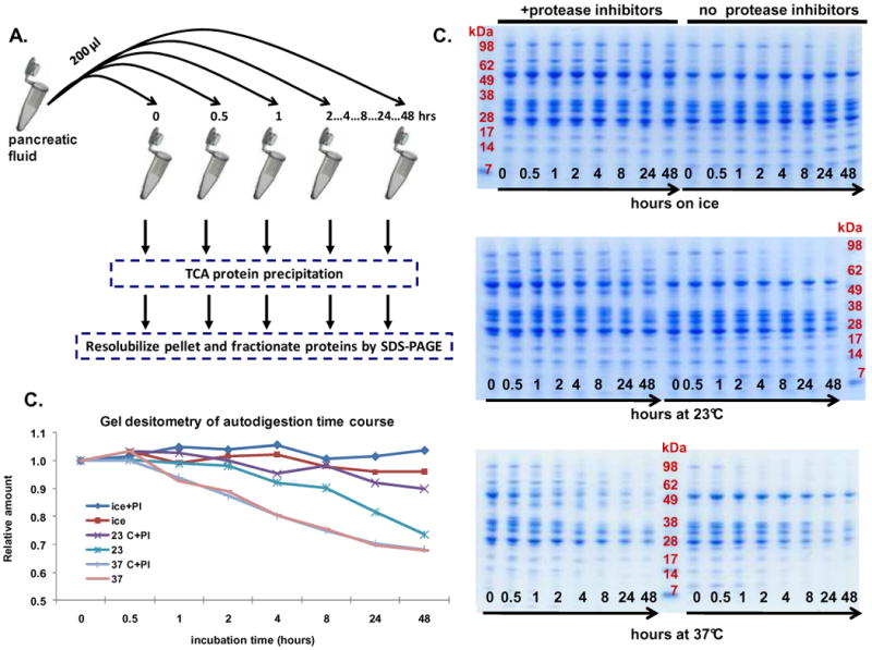
Figure 4. Experiment 2- Evaluation of temperature and time on pancreatic fluid auto-digestion.

A. Workflow of auto-digestion experiment. B. SDS-PAGE separation of samples incubated on ice (top), at 23°C (middle) and at 37°C (bottom). C. Quantitative gel densitometry measurements of entire gel lanes as determined using ImageJ software. Data points for each curve were normalized using the reading at the t=0 time point.
